By Admin
Content
A clinician dims the room lights and lifts a Wood's lamp toward a patient's forearm. A hypopigmented patch that barely registered in white light now shows a sharp, bright border. Diagnostic clarity like this depends on a single specification: the Wood's lamp wavelength. Choose the right peak, and fungal infections, pigment disorders, and porphyrin-related conditions become easier to screen. Choose the wrong one, and the fluorescence you expect may never appear. This article explains what Wood's lamp wavelength actually means, why 365 nm remains the clinical benchmark, and how 395 nm LED alternatives fit into a real dermatology workflow.
A Wood's lamp is a UV-A light source that passes through a filter designed to block almost all visible light. Robert Williams Wood introduced the original barium-sodium silicate glass more than a century ago, and the principle has not changed since. The classical description, repeated in JAMA and major dermatology references, is a band between 320 nm and 400 nm, centered around 365 nm.
Modern LED models have added new numbers to the market. Devices are sold with peak wavelengths of 365 nm, 375 nm, 385 nm, or 395 nm, and the medical literature now commonly cites 395 nm as a representative value. That figure is not wrong for a modern narrowband LED, but it describes a different optical situation from the instrument used to build most of the evidence base. A peak wavelength tells you where the lamp emits most strongly; it does not tell you how well it excites the molecules you are trying to visualize.
Fluorescence starts with absorption. A molecule gains a photon and quickly re-emits light at a longer wavelength. The fluorophores that matter in skin examination (collagen, elastin, NADH, porphyrins, and dermatophyte metabolites) absorb most efficiently in the mid-UV-A range. At 365 nm, they absorb well and emit visible light strong enough for the naked eye to read. At 395 nm, the absorption efficiency of several of these compounds drops noticeably, which means dimmer fluorescence and less contrast.
The existing evidence reinforces the point. A commonly cited review in the Journal of Clinical and Aesthetic Dermatology describes the 320-400 nm emission range with a 365 nm peak as the standard, while StatPearls references 395 nm for modern devices. The practical reality is that vitiligo borders, tinea capitis fluorescence, and porphyrin patterns were all characterized with a 365 nm source. If your goal is reproducible diagnostic work, matching that wavelength is the safest route.
The clinical literature and the device market converge on two practical peaks. The table below summarizes how they differ.
| Feature | 365 nm | 395 nm |
|---|---|---|
| Emission range | Broadband UV-A, 320-400 nm | Narrowband violet, ~395 nm |
| Typical source | Mercury vapor lamp + Wood's glass | Sealed LED |
| Fluorescence excitation | Strong for skin fluorophores | Moderate to weak for several targets |
| Visible violet glare | Minimal | Higher |
| Clinical evidence | Decades of published literature | Emerging, limited comparative data |
| Warm-up time | Several minutes | Instant |
| Device cost | Higher | Lower |
Both wavelengths can detect the major conditions: vitiligo, erythrasma, and dermatophytosis among them. The real question is how much diagnostic confidence you need. If your practice leans on precise pigment border assessment, 365 nm is the safer investment. If you need a lightweight, instant-on screening lamp for a busy general clinic, a 395 nm LED is defensible, provided you verify its output against known positive cases before relying on negative results.
Vitiligo is the classic Wood's lamp diagnosis. Depigmented patches appear as chalk-white areas with strikingly sharp borders because the absence of melanin removes competing absorption. Hypopigmented conditions such as pityriasis alba show softer, less defined edges. The same technique is also used to identify ash-leaf spots in tuberous sclerosis and to map the extent of melasma before starting treatment.
Microsporum canis, an ectothrix dermatophyte, produces fluorescent metabolites that glow apple-green under UV-A. Erythrasma from Corynebacterium minutissimum fluoresces coral-red, and Pseudomonas-infected wounds may show yellow-green. These colors are reliable screening cues, but a negative exam does not clear a fungus: Trichophyton species and several other common pathogens simply do not fluoresce, so Wood's lamp findings always need culture or microscopic confirmation.
Propionibacterium acnes generates porphyrins that emit orange-red fluorescence, which is why Wood's light is routinely used to map acne distribution. Urine fluorescence testing for porphyria cutanea tarda works on the same principle. These applications sit squarely in the excitation range of a 365 nm source and are noticeably weaker under 395 nm illumination.
Technique determines whether a wavelength advantage turns into a usable diagnosis. Follow this sequence:
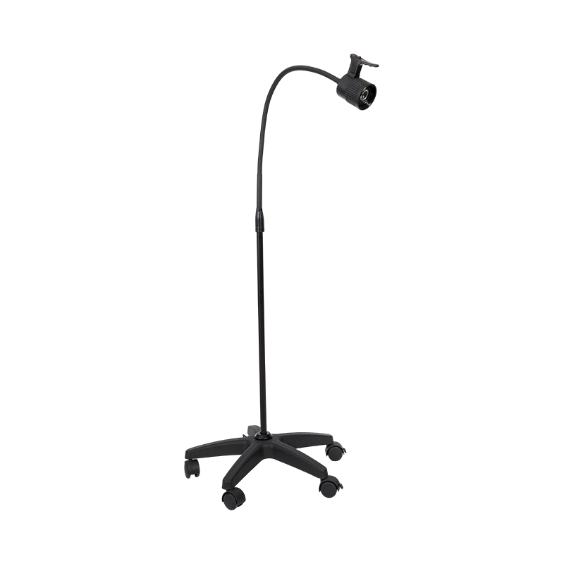
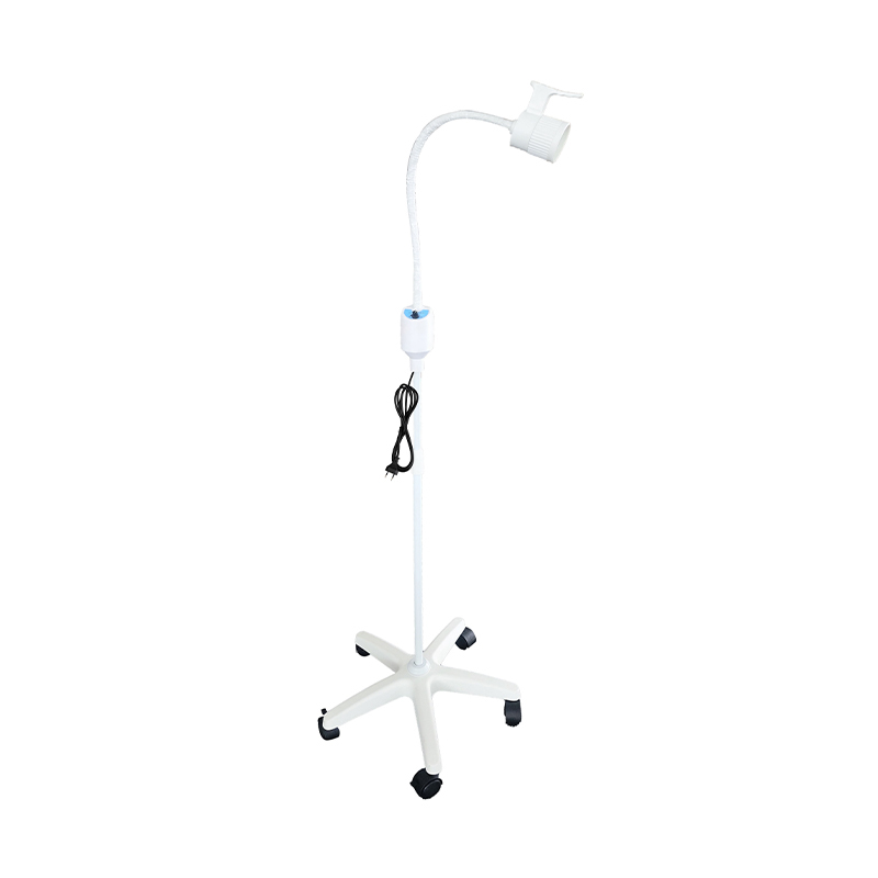
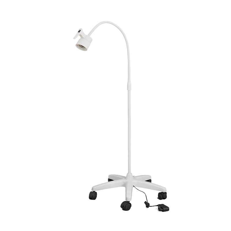
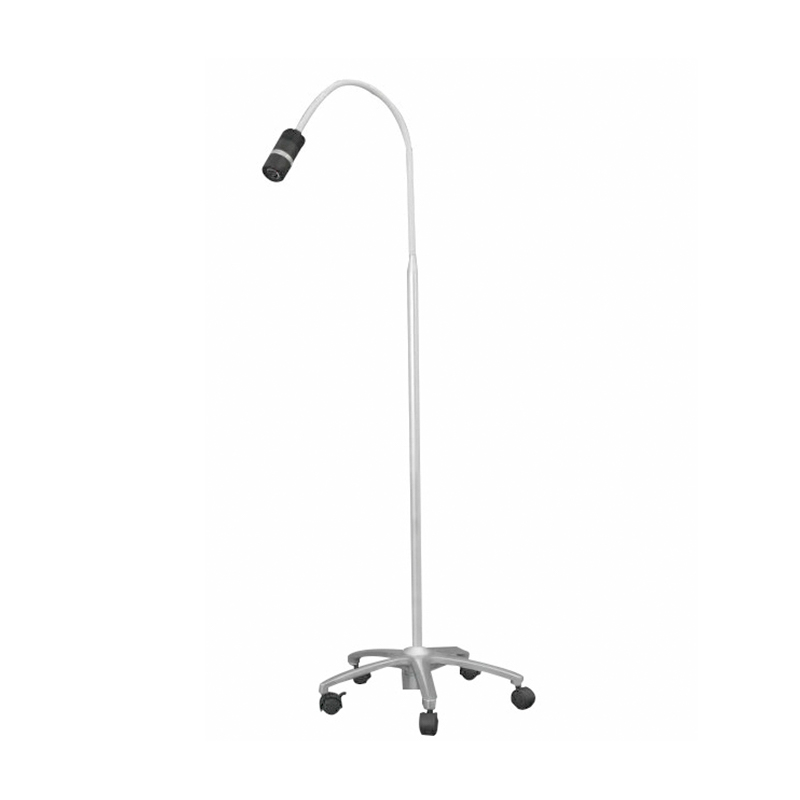
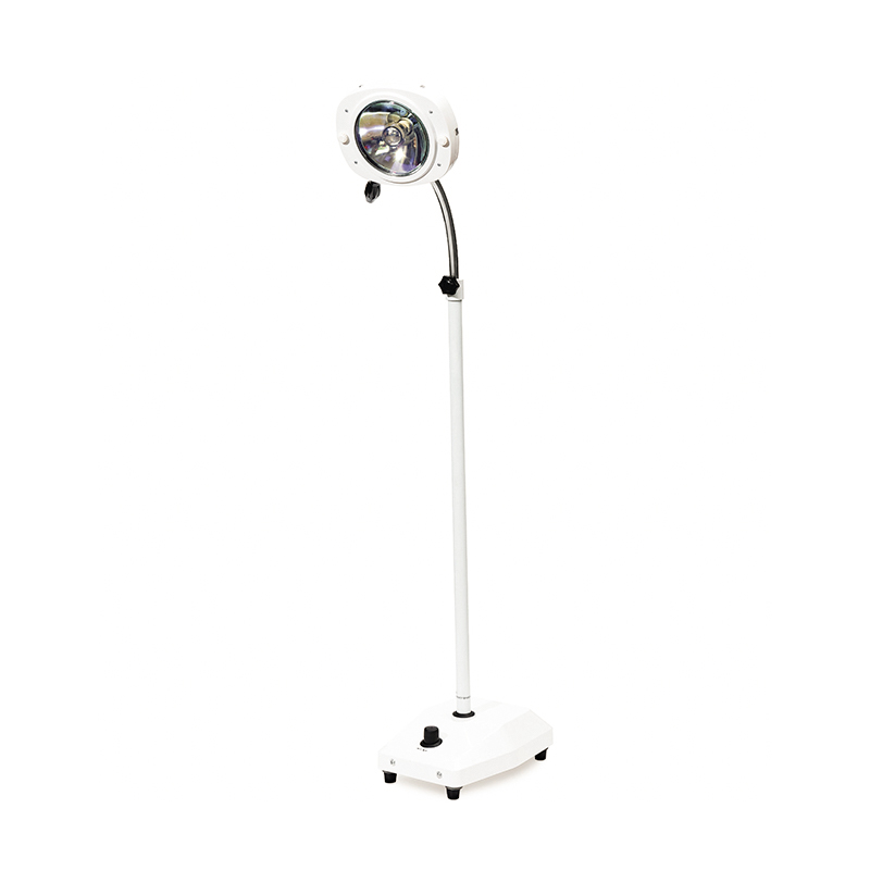
Ask the patient to remove creams, powders, or cosmetics beforehand because many topical products fluoresce on their own and produce false positives. After you identify a suspicious area, transfer the finding to shadow-free white light for marking, measurement, and photography. That is where a 12 W gooseneck LED examination lamp with adjustable spot size earns its place on the examination cart.
 Wholesale 12W Led Spot Size Adjustable Gooseneck Examination Lamp Suppliers, OEMJiangyin jianshifu Equipment Co.,Ltd is top China 12W Led Spot Size Adjustable Gooseneck Examination Lamp suppliers and OEM company, We s...View Product →
When you compare Wood's lamp suppliers, do not evaluate wavelength alone. Inspect the filter against the source, check the peak wavelength on the datasheet rather than the marketing copy, and decide whether you need a battery-operated unit for bedside work or a fixed unit for the treatment room. For fluorescence-based diagnosis, a 365 nm source with genuine Wood's glass remains the recommended combination. If budget pressure favors a 395 nm LED, accept it only as a screening instrument and keep that limitation documented in your protocols.
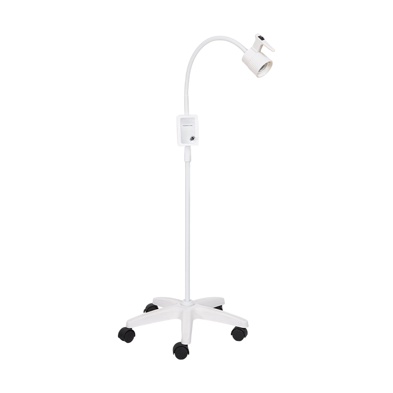
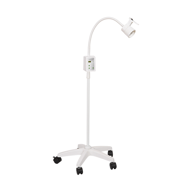
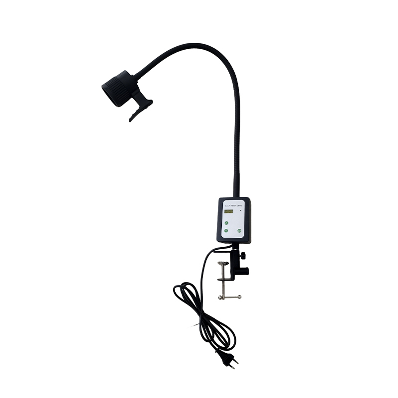
Planning a purchase is also a good time to review practical guidance on UV light equipment for healthcare settings so your choices align with current practice. And remember that the Wood's lamp is not the only lamp in the room. Routine skin checks, suture removal, and photography depend on a dependable white-light workhorse such as a compact 3 W LED examination lamp that mounts on a gooseneck without taking up counter space.
 Wholesale 3W LED Examination Lamp Suppliers, OEM Company - Jiangyin jianshifu EqJiangyin jianshifu Equipment Co.,Ltd is top China 3W LED Examination Lamp suppliers and OEM company, We specializes in wholesale 3W LED E...View Product →
UV-A is not the same as the UV-B that causes sunburn, but cumulative exposure is still worth respecting. Keep examinations brief, never point the lamp at the eyes, and do not hold the source close to the skin longer than necessary. The lamp is a screening aid, not a histopathology substitute. A confident examination is one that knows its own limits. Fluorescence narrows the differential, but the final diagnosis rests on clinical correlation and confirmatory testing.
The Wood's lamp wavelength is not a minor datasheet detail. It controls which fluorophores respond, how distinct the lesion margins look, and whether your findings match the evidence you rely on. Build your protocol around a purpose-fit source: 365 nm for diagnostic standard, 395 nm for cost-conscious screening, and give your exam room the white-light backup it needs for the rest of dermatologic care.
 We have specialised in medical devices for 32 years.
Contact Us
 No 48 Xiaokan Rd, Shizhuang town, Jiangyin, Jiangsu, China
 sara@jsf-qx.com
 +86-18915253915
 +86-18861287595
+86-18915253915
+86-18861287595
+86-18915253915
Copyright @ Jiangyin Jianshifu Equipment Co.,Ltd.
 Custom Disinfection Equipment Manufacturers
China Disinfection Equipment Factory
Custom Disinfection Equipment Manufacturers
China Disinfection Equipment Factory
The information provided on this website is intended for use only in countries and jurisdictions outside of the People's Republic of China.


 English
  عربى
عربى